Automatisches Perimeter
Automatic Perimeter
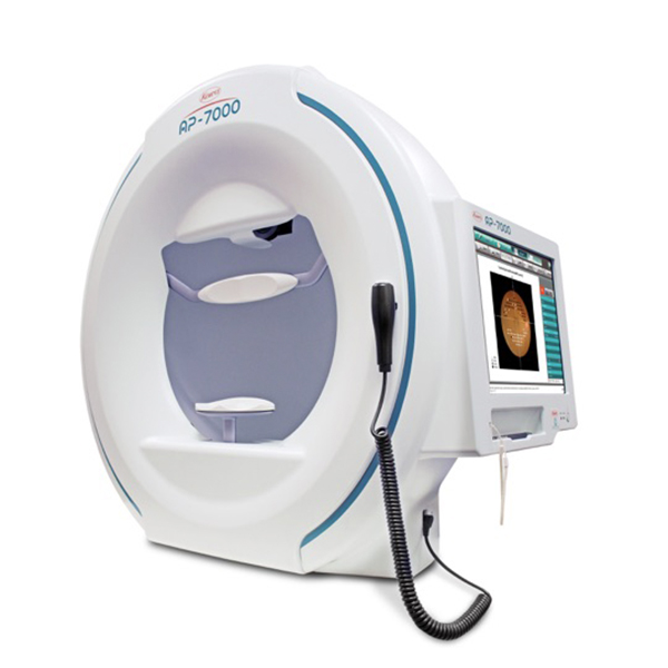
AP-7000
AP-7000
Das AP-7000 ist das Kowa-Perimeter der nächsten Generation mit Goldstandard-Schwellentest, das Ihnen zuverlässige und einheitliche Untersuchungsergebnisse des Gesichtsfelds Ihrer Patienten liefert.
- intuitive Bedienung
- umfassende Auswahl an Test- und Screeningstrategien
- Quick-Modi für Schwellenwerttest- und Screeningprogramme
- automatische Korrelation von Fundusaufnahmen mit der statischen Gesichtsfelduntersuchung
- einfache Bedienung über Touchscreen
- ergonomisches und kompaktes Design
- Unterstützung der Glaukom-Früherkennung
- Möglichkeit der Netzwerkanbindung für einen effizienten Arbeitsablauf
The AP-7000 is the next generation Kowa perimeter with Gold Standard Threshold Test that gives you reliable and consistent examination results of your patient's visual field.
- intuitive operation
- comprehensive selection of test and screening strategies
- quick modes for threshold test and screening programs
- automatic correlation of fundus images with static visual field examination
- Easy touchscreen operation
- ergonomic and compact design
- support for early glaucoma detection
- network connectivity for efficient workflow
- easy to use touchscreen operation
- easy to use and easy to use
Beschreibung
Description
Einheitliche und zuverlässige Untersuchungsergebnisse
Durch die umfassende normative Datenbank von Kowa, in der sowohl das zentrale, als auch das periphere Gesichtsfeld berücksichtigt werden, können Sie sich sicher sein, immer einheitliche und genaue Untersuchungsergebnisse zu erhalten.
Vielfältige Untersuchungsmöglichkeiten
In den Schwellenwertmodi ist die Untersuchung von Makula, Zentrum und Peripherie bis zu einem Winkel von 80° möglich, wohingegen in den Screeningmodi eine schnelle Beurteilung des Gesichtsfelds ermöglicht wird. Zur Verkürzung der Untersuchungszeit stehen sowohl für Schwellenwert- als auch für Screeningtests Quick-Modi zur Verfügung.
Verknüpfung der Perimetrie mit Fundusbildern von Funduskamera, OCT oder SLO
Die Daten der statischen Perimetrie können mit Fundusbildern aus Kameras, OCT und SLO verknüpft und spezielle Untersuchungslokalisationen auf der Retina definiert werden.
Einfache Bedienung, ergonomisches Design
Das ergonomische, kompakte Design des AP-7000 verbindet zusätzlichen Patientenkomfort mit Anwenderfreundlichkeit und fügt sich perfekt in die Praxis ein.
- Netzwerklösung
- vorbereitet für den Anschluss an ein Netzwerk mit integriertem PC
- einfache Netzwerkverbindung mit anderen Systemen zur Korrelation von Daten
- Export von Patientendaten, Untersuchungsergebnissen und Bilddateien an Ihr EMR-System
- einfaches Speichern und Ausdrucken von Gesichtsfeld-Plots
- Verbindung zur Kowa Netzhautkamera
Uniform and reliable diagnostic results
Thanks to Kowa's comprehensive normative database, which takes both central and peripheral visual fields into account, you can be sure that you will always receive consistent and accurate results.
Manifold examination options
In the threshold modes the examination of macula, center and periphery up to an angle of 80° is possible, whereas in the screening modes a quick evaluation of the visual field is possible. Quick modes are available for both threshold and screening tests to shorten the examination time.
linking perimetry with fundus images from fundus cameras, OCT or SLO
The data of static perimetry can be linked with fundus images from cameras, OCT and SLO and special examination locations can be defined on the retina.
Simple operation, ergonomic design
The ergonomic, compact design of the AP-7000 combines additional patient comfort with user-friendliness and fits perfectly into the practice.
- network solution
- prepared for connection to a network with integrated PC
- easy network connection to other systems for correlation of data
- export of patient data, examination results and image files to your EMR system
- easy saving and printing of visual field plots
- Link to the Kowa retinal camera
- easy connection to a network with other systems for correlation of data
- easy export of patient data, examination results and image files to your EMR system
- easy saving and printing of visual field plots
- Link to the Kowa retinal camera
- easy connection to your EMR system
- easy connection to a network with an integrated PC
- easy connection to other systems for correlation of data
- easy export of patient data, examination results and image files to your EMR system
- easy saving and printing of visual field plots
- easy connection to the Kowa retinal camera
Software
Optionale Viewer Software zur Bearbeitung der Daten am Computer
Optional viewer software for editing the data on the computer